

Amelogenesis Imperfecta (AI) disrupts the composition of enamel in teeth. The condition can be x–linked, autosomal recessive or dominant. AI can affect both permanent and primary teeth. Clinical presentations of AI can include sensitivity, surface roughness, and discoloration. Diffuse pitting or grooves are caused by defective matrix deposition, showing up as reduced enamel thickness. Most often AI presents with significant surface wear, potentially associated with loss of vertical dimension of the patient. Due to the brittle surface enamel of teeth, usually a multidisciplinary approach is needed, and treatment should be planned during childhood when symptoms are first noticed.
Treatment of dental conditions when presenting alongside AI can be challenging. However, when balancing underlying conditions and patient motivations, we can achieve a very stable and healthy long-term result.
Case Presentation
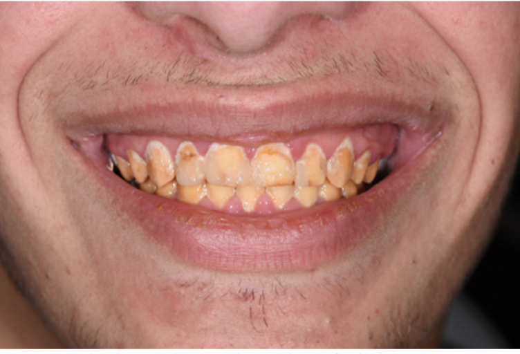
A 17-year old male patient presents when referred by the orthodontist. The patient has finished 4 years of orthodontic treatment. The patient presents with Amelogensis Imperfecta, which is also present in his sister and mother. The patient’s chief concern is to restore esthetics.

Clinical Findings
- Dark yellow and mottled teeth, which are very brittle. The surface enamel can be easily removed with hand scalers.
- All teeth show short clinical crowns, and significant evidence of incisal/ occlusal wear is evident.
- Periodontal health and oral hygiene are poor, but the gingival levels are acceptable.
- There is no display of teeth when lips are at rest.
- Radiographic examination shows good bone levels with no periapical pathologies.
Treatment
- To build a strong and healthy foundation for the final restorations, the patient was referred to a periodontist. Scaling and root planning was carried out, along with a 3-month recall schedule.
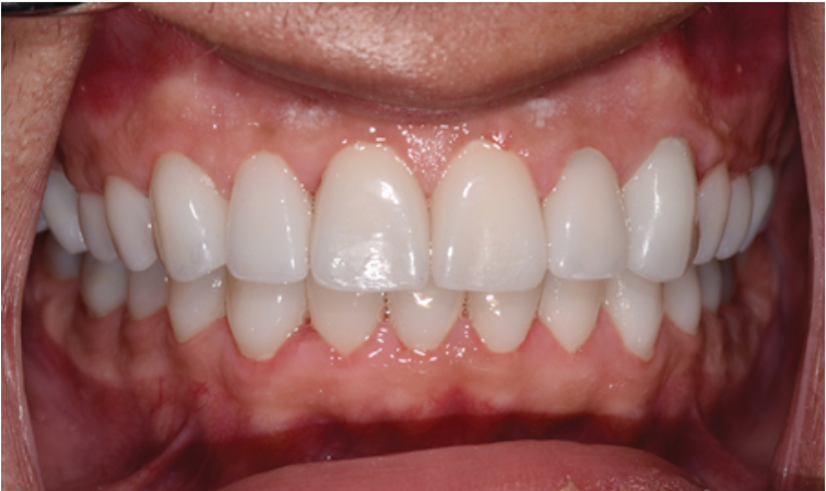
- After restoring periodontal health, a restorative treatment plan of full coverage monolithic full contour zirconia crowns was developed. This decision was made in order to:
- Restore the lost vertical dimension of teeth by 2 mm.
- Protect against further enamel wear due to a combination of parafunction and brittle/ soft enamel.
- Following preparation of the teeth, temporary crowns were made using a diagnostic wax up. The temporary crowns were kept for 2 months. This was done to allow for periodontal healing, patient adjustment to new phonetics, adjustment to new function, and prevention of any sensitivity.
- The final full contour monolithic zirconia crowns were cemented with a resin based luting cement.
- An occlusal appliance was also made following end of treatment, to further protect against parafunction.

This case was completed and published in Oral Health by Dr. Aragon (Prosthodontist in London, Canada).