
")
The final esthetic outcome and longevity of veneers depends on the tooth preparation. Excessive reduction can compromise bond strength (due to exposed dentin), cause sensitivity and limit possible treatment options for the patient in the future. The goal of veneer preparations is to be maintained within the enamel layer. Conservative tooth preparation can give high fracture resistance with the use of resin cements.
This is where diagnostic wax ups, a clear matrix and a putty matrix can be extremely helpful.
A diagnostic wax up can be used to determine ideal tooth sizes, occlusion, and the working space available. Critical to patient communication, diagnostic wax ups can be transferred into the mouth with temporary self-curing resins, to give the patient a clear vision of their future smile.
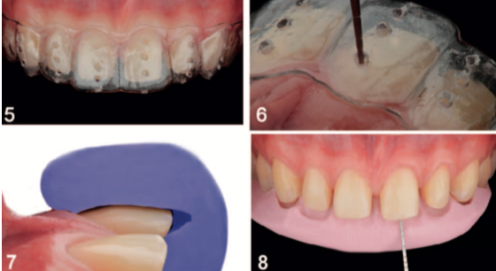
A clear thermoplastic matrix, made with a vacuum machine can give an overall evaluation of the preparation of the teeth. This can be further enhanced with perforation in different areas of the matrix and measurement of the reduction with a perio-probe.
A putty matrix provides reduction measurements in specific areas of a tooth, such as the facial or incisal. It gives the clinician a clear 3-D view of their current position in their reduction roadmap.
Here we present a case report outlining the use of these reduction guides.
Case Presentation by Jurado et al.
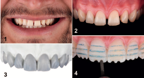
- A 31- year old male presents to the dental office with a chief concern of closing interproximal spaces and improving his smile.
Clinical Exam
- Interproximal spacing between all maxillary anterior teeth – canine to canine.
- Uneven incisal edges from upper right lateral incisor to upper left lateral incisor.
- Evidence of wear on incisal edges of all maxillary anterior teeth.
- Large buccal dark triangles (smile corridor) were also evident when the patient smiled.
Treatment Plan
- Orthodontics and veneer restorations were presented as treatment options. As the patient did not want to go through with orthodontic treatment, veneer restorations were chosen as the treatment plan.
- Maxillary canine-to-canine porcelain veneers were planned to close anterior spacing, create even incisal edges and brighten the smile.
- Ceramic veneers were planned for upper right and left first premolars to decrease the buccal dark triangles (smile corridor).
Treatment
- The laboratory fabricated a diagnostic wax up of the proposed final restoration. A putty matrix and a thermoplastic clear matrix were made with a vacuum machine. In the clear matrix, three small perforations were made on the facial surfaces of each tooth, in order to be able to measure the reductions with a perio-probe.
- A mock-up was placed in the patient’s mouth with self-curing temporary resin material. The patient and clinical were happy with proposed final restoration.
- Horizontal and vertical depth grooves were cut into the teeth with a round diamond bur.
- Tooth reduction was done with the aid of the clear matrix and repeatedly measured with a perio-probe.
- For a better visualization of the incisal and facial reductions, the putty matrix was repeatedly fitted.
- The prepared teeth were sandblasted with water and 20-micron aluminum oxide particles.
- The final impression was taken with double cord placement, and light-body/ heavy-body polyvinylsiloxane impression material.
- Feldspathic veneers were chosen to match the high esthetic demands of the case and to be able to produce thin veneers. The ceramic veneers to be placed on the premolars were made out of feldspathic porcelain.
- For cementing the veneers, rubber dam isolation was used. The internal surface of the veneers were treated with hydrofluoric acid and silane. The tooth surfaces were treated with 32% phosphoric acid. Optibond primer and adhesive were applied and light cured. Variolink light colored cement was applied to the veneers, placed on the teeth, excess cemented removed and light cured. Occlusion, excursive and protrusive movements were checked.
- An occlusal guard was also fabricated to prevent any damage to the veneers and natural teeth.


By using preparation guides, we can make sure that our tooth preparations are as conservative as possible. A clear matrix gives a good overall perspective of the reductions made on a tooth, while a putty matrix allows us to evaluate the incisal and facial surface specifically.