

The following presentation is the third and final case series on the management of macrodontia of fused anterior maxillary incisors.
Part 3
As part of continued discussion on anterior maxillary tooth fusion we’ll be presenting our 3rd and final treatment case series. Thus far we’ve reviewed a conservative approach in case 1, with the authors choosing to defer surgical and endodontic treatment on a vital maturing adolescent fused maxillary incisors. We changed scopes and presented an approach in treatment of necrotic fused teeth of 20-year-old patient, following a regiment that entailed multidisciplinary intervention for case 2. As we’ve seen so far treatment of fused teeth can be complicated; however, with diligent planning and communication great outcomes can be achieved and maintained over time. Tune in for the third and final case below:
Case 3:
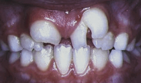
Patient: 9-year-old girl
CC: Esthetic concern of maxillary central incisor
RMH: Non contributory dental, medical and familial history
IOE:
- Fusion of central left incisor with supernumerary tooth
- Clinically regular tooth count for arch
- Diastema between fused teeth and right central incisor
- Right and left central incisors measured at 10mm, 15mm mesiodistally
- Skeletal class 2 Div 1
- Normal pulp vitality
Radiographic Exam:
- Radiographic exam revealed fused tooth had two separate root canals and two distinct roots
- Normal periapical status noted
Treatment:
- Full thickness buccolingual flap and sectioning of the mesial portion of the fused tooth, plane of sectioning termination located subgingivally
- Direct pulp cap with MTA at root midline, restored with direct composite
- Orthodontic treatment initiated upon asymptomatic 12 week follow up
- Left maxillary incisor proved vital upon 10-year follow up.
Final Thoughts
Surgical treatment may be considered depending on root morphology. Pulp capping with MTA may be viable option in preserving a healthy pulp tissue. MTA releases Ca++ resulting in formation of hydroxyappetite and is a promising material when used for capping after partial or total pulpotomy as noted with patient left central incisior vital 10 years following treatment. Implant is not a recommended treatment option for a child or adolescent during maturation phase. CBCT can serve as a vital tool, especially in establishing root configuration.
This case was completed by Dr. Nelly Steinbock et al. view citation for full publication
DOI:https://doi.org/10.1016/j.joen.2013.12.004