

The following presentation is part of three case series on management of macrodontia of fused anterior maxillary incisors. Tune in each week as we discuss each unique scenario.
Part 1
Macrodontia of anterior teeth may be an isolated condition or occur as a result of fusion, gemination, or concorcense of teeth.
Just to refresh:
- Fusion of teeth occurs between two adjacent tooth germs, involving the confluence of dentin.
- The fusion may be complete or incomplete depending on the developmental stage at the time of the union.
- Pulp chambers and canals may be joined or separate depending on the time of fusion.
- Fusion that occurs prior to the calcification stage will result in complete union.
- There is one tooth fewer than the normal count for the arch, however, fusion with supernumerary teeth may occur resulting in a normal count for the arch.
- Thought to be resultant of physical forces producing the necrosing of intervening tissue causing contact between enamel organ and dental papilla.
- Gemination occurs when one tooth bud attempts to divide, resulting in a bifid crown and single root.
- Tooth count for the arch is normal.
- Concorecense is the union of two already formed teeth via cementum.
These anomalies are rare, observed in less than 1% of the population, often affecting anterior teeth and occur in both jaws. Management of macrodontia of teeth with fusion, gemination, and or concorencense presents a unique challenge because they often present with anomalous size and shape of crown, and cause esthetic, periodontal, orthodontic problems for the patient clinically. These cases often require multidisciplinary management in achieving success. Tune in below for the discussion of the following three cases:
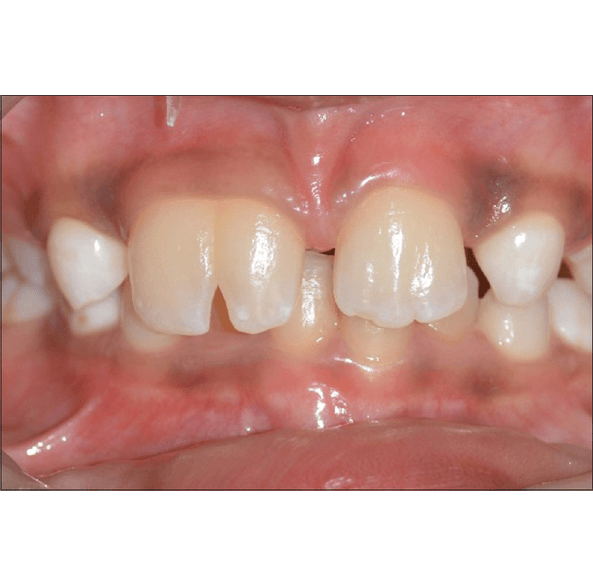
| Case 1 Patient: 10-year-old boy presents to clinic CC: Poor esthetic of upper right incisor RMH: Non contributory dental, medical and familial history IOE:Macrodontia of maxillary right and left incisors measured at 16.5, 12.5mm respectivelyClinically missing lateral incisorsComplete fusion of left maxillary incisorsIncomplete fusion of right maxillary incisorsBifurcation grooves noted buccalingually of the right maxillary incisorsVitality testing of both incisors and surrounding periodontal tissue WNLMixed dentitionRadiographic Exam:CBCT revealed two roots, two distinct but connect root canals of the left incisorsRight incisor had one root and one canalTreatment:0.5 mm veneer preparation of the right maxillary incisorDigital mock up of veneer fabricated with Adobe Photoshop and Corel Draw software to mimic the shape of the left central incisor and dictate appropriate margins and shape of the lateral incisorIPS emax veneer fabricated and cemented with dual cure resin luting rely X cement, pink ceramic utilized in mimicking gingiva |
| Final Thoughts Veneering can be an appropriate treatment option in order to gain the desired esthetics and maintain vitality of the pulp and adequate tooth structure in a 10-year-old child. Orthodontic treatment can be an appropriate option in order to gain space for the missing left lateral incisor. Regular recall appointments are necessary to avoid any periodontal or caries processes from taking place in the lingual groove. Right maxillary fused incisor proved asymptomatic upon 2-year follow up. |
This case was completed by Dr.Godvin Clovis Da Costa et al. view citation for full publication
DOI: 10.4103/ccd.ccd_1090_16