

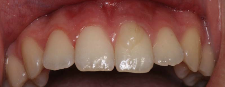
A 16-year-old girl presented to the clinic with a fistula on the apical third of her upper left central incisor. During clinical examination, a tubercle was seen on the buccal cervical third of the tooth.
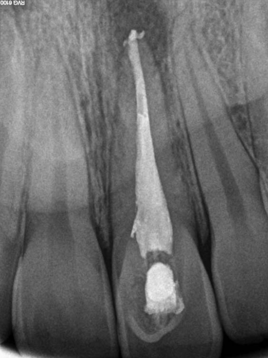
Radiographic and clinical examinations provided a diagnosis of apical chronic supurative periodontitis. Root canal therapy (RCT) of the upper left central incisor was indicated.
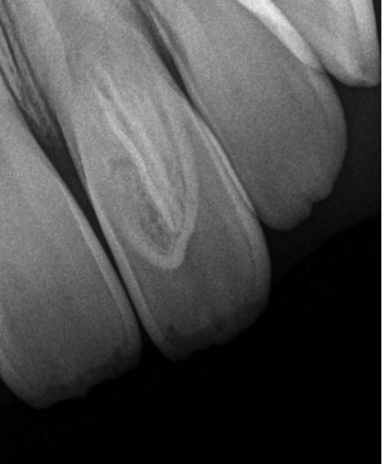
The tubercle brought upon by the dens evaginatus of the tooth presented an unusual pulpal anatomy. Unlike supplementary cusps, such as the cusp of Carabelli, dens evaginatus tubercles can present with pulpal extension 70% of the time. Therefore, a CBCT was taken in order to fully understand the pulpal configuration. Sagittal cuts showed the presence of pulp tissue inside the buccal tubercle. Therefore, a typical access cavity could not be used for the RCT.

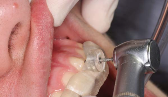
It was decided that a splint would be used to guide the preparation of the access cavity. The same software used for guided implant placements (SIMPLANT) was used to fabricate a guided splint for accurately determining the shape and extent of the access cavity needed.
Rubber dam isolation was used after access cavity preparation. Working length was determined with Root ZX electronic apex locator and the canals were prepared with Protaper Next (Dentsply) with the use of sodium hypochlorite irrigation. Finally the canals were filled with a warm vertical condensation technique.
For the final restoration, the tooth was prepared for veneer placement to eliminate the buccal defect of the tooth.
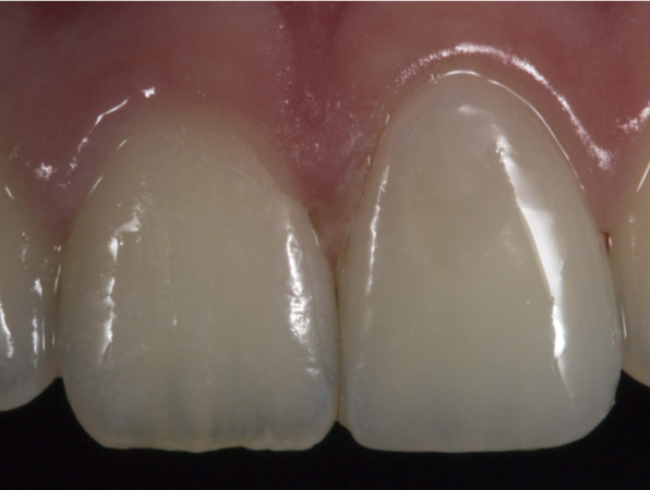
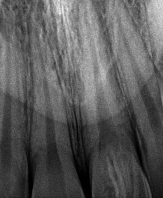
Follow up CBCT was performed in one year, and full apical healing was observed.
Dens evaginatus is thought to develop from unusual proliferations of the enamel epithelium during the bell stage of tooth development. As mentioned, the majority of dens evaginatus cases present with extensions of the pulp tissue into the accessory cusp, with a very thin layer of enamel covering that portion of the pulp. The patient can present with severe sensitivity, and potential pulp exposure from even slight attrition, enamel wear or cracks during regular function. Dens evaginatus can affect all teeth, but is mostly seen in premolars, presenting bilaterally and symmetrically.
This case was completed and reported by Jesu s Mena-A lvarez, DDS, PhD, Cristina Rico-Romano, DDS, PhD, Ana Bele n Lobo-Galindo, DDS, lvaro Zubizarreta-Macho, DDS, PhD.