

Patient Presentation
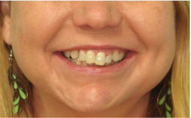
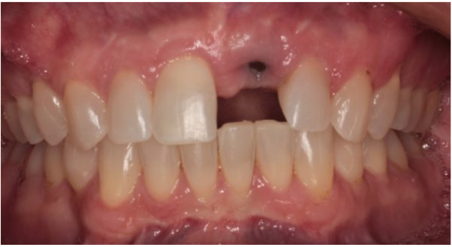
A patient presents to the orthodontic office with a chief complaint of straightening her upper and lower front teeth. She is particularly concerned about the vertical height of the two upper central incisors, which also cause an unbalanced upper lip position at rest.
Dental history
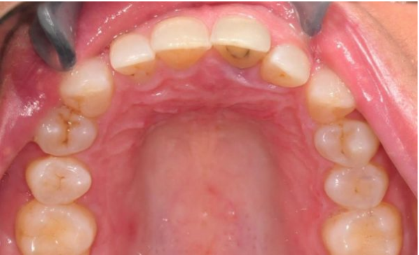
- Previous root canal treatment of upper left central incisor.
- Regular hygiene and recall appointments.
Treatment
The comprehensive treatment plan required a collaboration between orthodontics, periodontics, endodontics, and restorative dentistry.
- Evaluation of the previous endodontic treatment of the upper left central incisor warranted retreatment. During evaluation, pathology was also evident with the upper left lateral incisor, which was also root canal treated at the same time. During the procedure, the upper left central suffered an apical vertical root fracture. This was treated with apicoectomy, preserving the proximal 1/3 of the root. This was done to preserve the bone in that space.
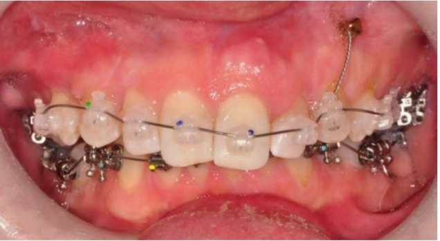
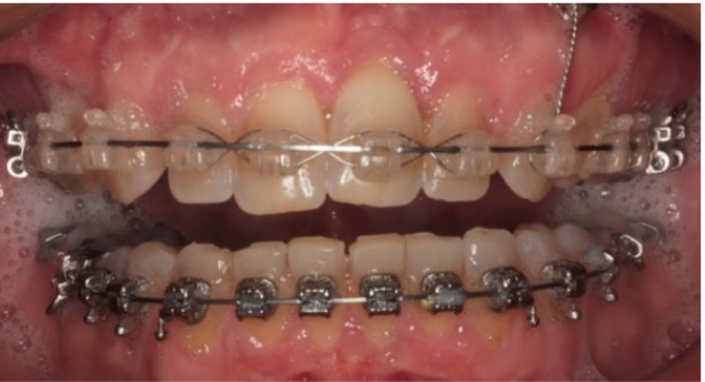
- Orthodontic treatment was initiated to balance the smile and specifically to correct the vertical discrepancies of the upper anterior region. Ormco Damon D2 and Damon Clear2 brackets were placed, along with TAD placement in the anterior region of the maxilla (Ormo VectorTAS 6mm TAD). Following two years of orthodontic treatment, the final desired tooth positions were achieved. Occlusal adjustments were done and upper and lower permanent retainers were placed.
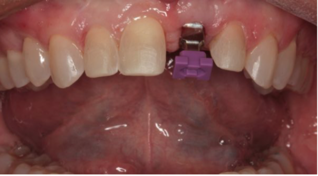
The upper left central incisor was extracted and an immediate implant was placed (Straumann bone level RC size 4.1 mm x 10 mm). The healing abutment was RC 5 mm x 4 mm. During implant placement, a residual space was maintained in order to have working room during the restorative phase, in order to achieve golden proportions.
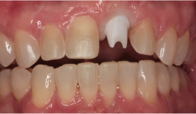
The restorative treatment included placement of an implant crown to restore the upper left central, and porcelain veneer placement from premolar to premolar on the maxilla (1M1 Vita 3D shade guide). The teeth were prepared with 0.5mm facial reduction, 1.5 mm incisal reduction, with chamfer margins at the gingiva and elbow margins at the proximal contacts. Provisional restorations were made with bisacrylic and retained with mechanical retention.
To restore the implant, a zirconia oxide abutment was fabricated. EMax implant crown and veneers were fabricated for the final restoration. The internal etched surface of the eMax crown and veneers were treated with silane primer (Bisco) for 20 seconds and air-dried. The crown and veneers were inserted with self-curing luting composite (Choice2, Bisco).
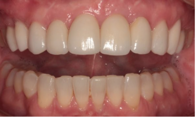
Planning the vertical position of the smile prior to initiation of treatment is critical for a case like this. Multidisciplinary collaboration and communication was essential.
This case was completed by Ross W. Nash, DDS (North Carolina) and Mark Allen, DDS (North Carolina) and also published in Oral Health.