
")
Case Report:
18 months after the initial cementation of a 3 unit fixed partial denture (FPD), a 48-year old woman returned to the office due to fracturing of the same FPD. Records showed that the FPD was made from a zirconia framework and veneered porcelain.
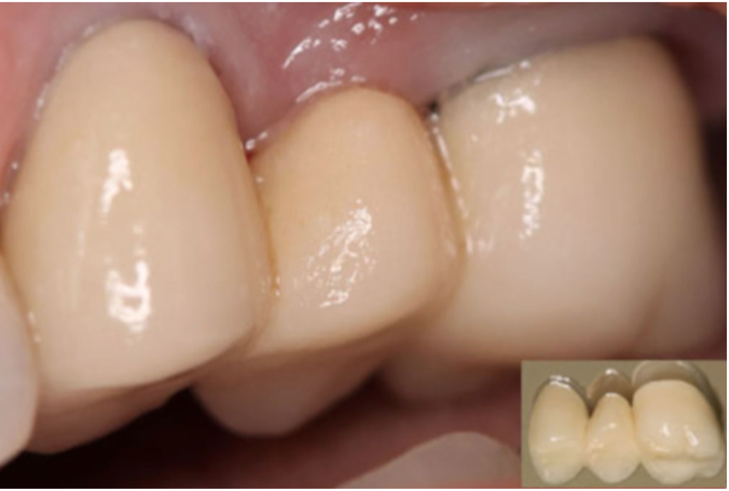
The patient presented with the fractured portion of the FPD in hand. Intraoral examination showed that the fracture occurred on the bucco-cervical portion of the pontic.
Repair attempt #1:
As the fractured portion of the FPD was available, the first attempt at repair was to re-cement the fractured portion to the rest of the bridge.
- The contacting surfaces of the broken fragment and FPD were etched with 10% hydrofluoric acid gel for 1 minute, rinsed and air-dried.
- Both surfaces were silinated for 60 seconds.
- Both surfaces were bonded with resin adhesive (Single Bond Universal) and light cured for 15 seconds.
- RelyX U200, a light cured resin cement, was placed on both surfaces, and the fragment was placed on the original pontic site. Excess cement was removed, and the cement was light activated for 40 seconds.

Repair attempt #2:
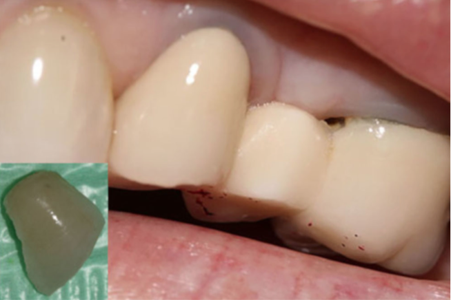
To the dismay of both the patient and the dentist, the patient returned almost 2 weeks later, for the same problem. However, this time, the fractured portion was lost.
This time, the only option was to repair the site with resin composite.
- The fracture site was beveled with a fine diamond bur.
- The fractured surface was etched with 10% hydrofluoric acid gel for 1 minute, rinsed and air-dried.
- The surface was silinated for 60 seconds.
- The surface was bonded with resin adhesive (Single Bond Universal) and light cured for 15 seconds.
- Composite resin (Filtek Z350) was applied in sequential 2-mm increments, and each light-cured for 20 seconds.
The final restoration was shaped and polished with extra-fine diamond burs and woolen felt burs with diamond paste.
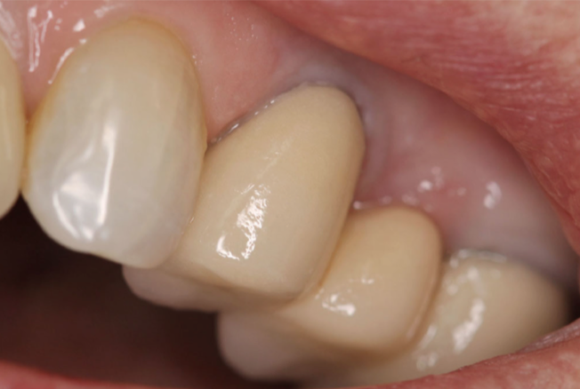
| The second attempt at repair has remained stable for 5 years and counting. So why the difference between the two clinical methods of repair? Resin composite is a lower elastic modulus material than the porcelain fragment. Therefore, the composite can absorb the energy of masticatory forces and distribute the stress in broader areas of the resin. Furthermore, the fractured pontic area was beveled first, in turn increasing the bonding area and the bond strength. The surface pre-treatment of the pontic was the most important part of the repair protocol – improving bonding surface area and using bifunctional agent silane. Most failures of the FPDs occur in the cervical areas of the pontics, as tensile stresses are concentrated in the cervical areas of pontics and connectors. Research has shown that resin composite repair of metal-ceramic restorations has an 89% survival rate. The next time your porcelain restoration chips – stay calm, cool and silinate. |
| Today’s Morning Huddle was reported by Dr. Potira Dalques Meirelles. |