

The following presentation is part of three case series on management of macrodontia of fused anterior maxillary incisors. Tune in each week as we discuss each unique scenario.
Part 2
As we discussed in our last week’s article, fusion of teeth occurs between two adjacent tooth germs, making physical contact and attempting to merge into a single tooth. Macrodontia of fused anterior teeth may be an isolated condition or occur as result of hypervitaminosis A, viral infection during pregnancy, use of thalidomide, osteopetrosis, chondroectodermal dysplasia, trauma and other environmental factors. The resulting fusion may be complete or incomplete yielding either separate or joint pulp chambers and canals depending on the time of fusion. Tooth count for the arch is one less than normal barring presence of supernumerary tooth. These cases are rare occurring in less than 1% of the general population and therefore treating them requires:
Attention to detail in case analysis of patient with diligent IOE and EOE, and review of radigoraphs, intraoral and other imaging modalities such as CBCTTreatment planning tailored to patient needs considering age and level maturation of patientInterdisciplinary communication and management of treatment.Communication with patient in gaining trust, acceptance and compliance of treatment is critical as such cases require intensive management and are often longer in duration.Adequate follow up and maintenance is an important aspect of longevity of health.
| Case 2: Patient: 20-year-old Caucasian male CC: Anomaly of his upper central incisor and excessive diastema RMH: Non contributory dental, medical and familial history IOE: Fusion of the two central incisors. Clinically missing tooth with regard to regular count for arch. Diastema between fused teeth and left lateral incisor. Dental misalignment. Bifurcation grooves noted buccalingually of the fused teeth extending 2mm subgingivally. Vitality testing were negative for fused central incisors and right lateral incisor Radiographic Exam: Radiographic exam revealed fused tooth had two separate root canals and two distinct roots. Periapical lesions of the fused central incisors and right lateral incisor noted. Treatment: Endodontic treatment of the necrotic fused central incisors and right lateral incisor. Endodontic apicoectomy surgery and retrograde obturation with reinforced zinc oxide-eugenol cement completed 6 months following initial endo treatment due to a persisting periapical radioleucency. Surgical separation of fused teeth in conjunction with initiation of orthodontic treatment after complete healing of periapex (6 months after apicoectomy)Frenulectomy completed. Appropriate positioning of teeth obtained after 9 months Prosthodontic preparation followed by definitive placement of anterior ceramic crowns. | 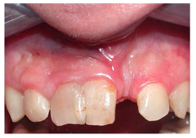  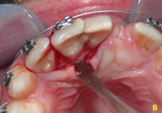 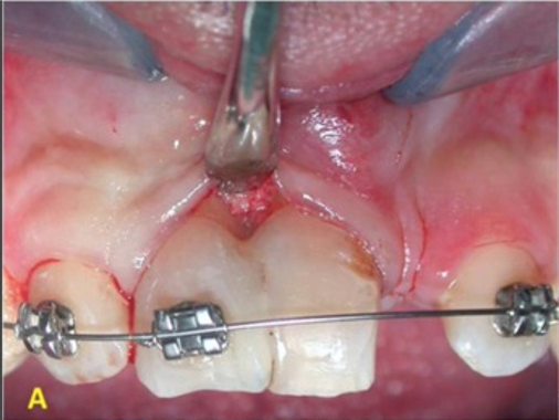 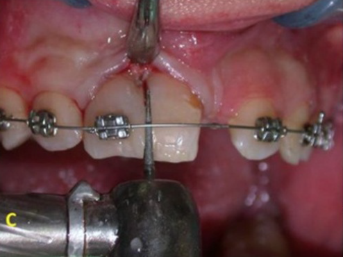 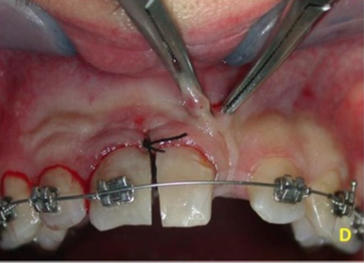 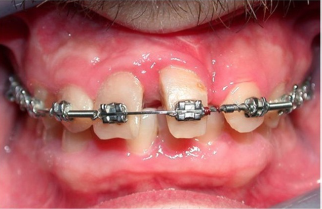 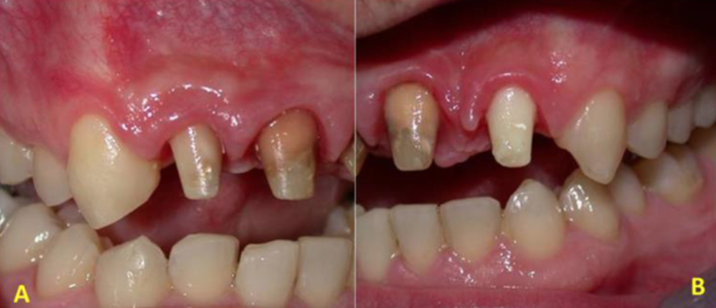 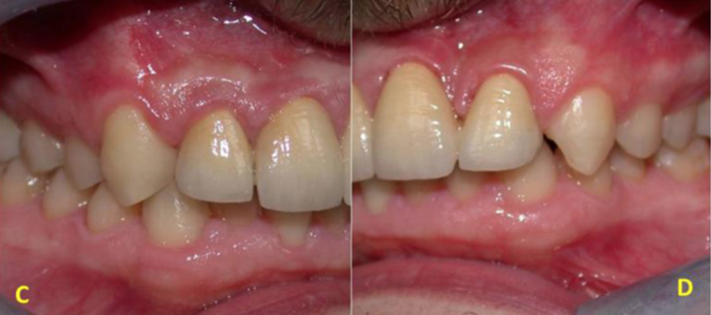 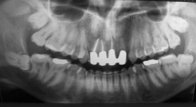 |
Final Thoughts
The prognosis of fused teeth is dependent on the area of connection of the two teeth, root development, age of the patient and their compliance with treatment protocol, pulp chamber and canal morphology. Such cases often require interdisciplinary treatment and communication for success.
This case presented by Gilberto Sammartino et al.
DOI: 10.1186/1752-1947-8-398