")
TMJ dislocations can be horrifying for our patients. A full TMJ luxation is when the condyle translates anteriorly of the articular eminence, and does not return to the fossa. This can occur after excessive mouth opening in yawning, eating or dental procedures. Patients present with a wide-open mouth, in a protruded position (during bi-lateral luxation) or lateral position to the non-affected side (during unilateral luxation).
The first treatment is the manual repositioning of the condylar head into the fossa by a clinician. Most of the time, the problem is solved. However, some patients with a steep articular eminence, weak capsule and ligament laxity are at a risk of having recurrent or chronic TMJ luxation events.
For patients with recurrent TMJ luxations, there are various non-surgical and surgical procedures to aid in prevention of future episodes. Initially we can start with jaw exercises to strength the muscles of the TMJ. Furthermore, we can perform injections of sclerosing or proliferant solutions and autologous blood into the TMJ capsule. There has also been much research done on injections of the botulism toxin in to the masseter or pterygoid muscles to decrease hyperactivity. For surgical procedures we can augment the articular eminence, perform lateral pterygoid myotomy or condylectomy.
Autologous Blood Injections
Autologous blood injections have shown the best scientific support for treatment of recurrent TMJ luxations. Since it is a non-surgical procedure, it is without all the complications of an inpatient surgical procedure using general anesthesia.
This procedure involves the injection of the patient’s own blood into the superior joint space and pericapsular tissues. With this procedure, the joint is first distended. Within a few hours, inflammatory mediators released by the platelets cause adjacent blood vessels to dilate and leak plasma, inducing further swelling of the area. In a few days following injection, organized blood vessels and loose fibrous tissues begin to form. As these tissues mature, joint stiffness is seen and a permanent limitation of the movement of the joint results. Due to this limitation in the range of motion of the TMJ, it is critical that the patient also undergo physical therapy to maintain range of motion. Autologous blood injections have shown 80% success rate for the treatment of recurrent TMJ luxations, providing a safe and cost effective treatment.
The Procedure
- The area overlying the TMJ is disinfected with an antiseptic solution.
- Local anesthesia is given to the auriculotemporal nerve.
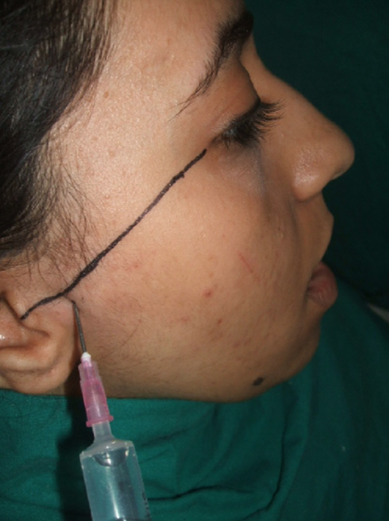
- The articular fossa is located 10mm anterior to the tragus of the ear and 2mm inferior to the tragal-canthal line (Figure 1).
- The joint space is flushed with saline, with the mouth in the open position, using an 18-gauge needle (Figure 1).
- The patient is asked to open and close several times to expel the fluid with the same injection needle.
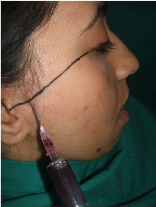
- Autologous blood is withdrawn from the patient anticubital fossa.
- 2 mL of blood injected into the superior joint space, and 1mL can be injected into the pericapsular tissues (Figure 2).
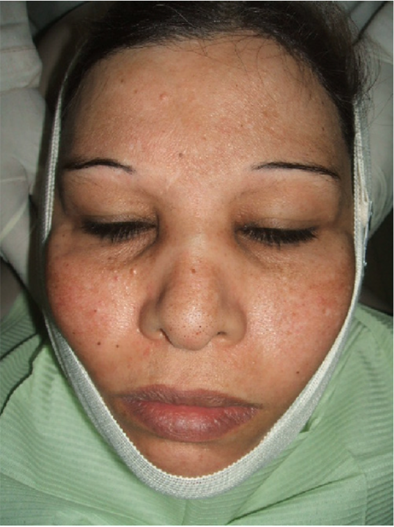
- An elastic bandage is applied over the TMJ and left for 24 hours (Figure 2).
- Patients are instructed to restrict in mouth opening and eat soft foods for seven days.
- Antibiotics and anti-inflammatory medications such ibuprofen are to be prescribed.
- Patients are followed up 2 weeks, 4 weeks, 3 months, and 6 months.